Do grew up and went to college in the Netherlands, where she received degrees in psychology and neuroscience. She is currently a neuroscience graduate student in the United States. Her interest is focused on understanding the neural substrates that underlie normal and abnormal brain functioning. She uses state of the art imaging methods, like magnetic resonance imaging (MRI) and diffusion tensor imaging (DTI) to examine alterations in white-matter structure. More specifically she is interested in investigating the connectivity that underlies affective processing in the brain. On the side she is also interested in the intersection between tech and health, and wants to develop novel tools to investigate and forecast healthy behaviors.
Studying connectivity in the brain is an integral part of understanding normal and abnormal behavior. This presentation will give an overview of the importance of researching the connectivity that underlies the neurobiology of risk factors for developing anxiety disorders. It is well know that the amygdala is important during fear processing, but a lot less is know about the involvement of the extended amygdala. A structure that will be more closely investigated in our research. This presentation is a reduced version of the original as presented at SfN 2012 that also included unpublished data.
Click image for presentation.
Diffusion Tensor Imaging (DTI) is a cutting edge imaging technique that provides quantitative information with which to visualize and study connectivity of neural pathways. A growing number of studies are now collecting DTI scans which necessitates knowledge of DTI processing steps and tools.
This presentation will give an overview of preprocessing steps like distortion correction and tensor estimation, discuss choices that can be made regarding whole brain versus tract specific analysis, and what normalization and visualization tools are available.
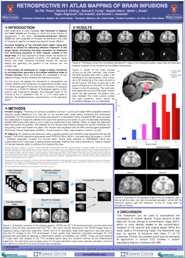
New treatments of brain diseases like Parkinson's disease are focusing on direct and local delivery of
therapeutics in the brain using Convection-enhanced delivery (CED). Accurate targeting of the intended brain region using this
method is critical for delivering effective treatment in the target tissue. In this project we developed an image analysis
framework to retrospectively generate a normalized statistical atlas of infusion studies.
This movie shows a colorful 3D rendering of a non human primate brain. Data from the Kalin lab, Department of Psychiatry, University of Wisconsin - Madison.
This movie shows a colorful 3D rendering of some of the fiber tracts in my brain. It shows the Corpus Callosum (CC), overlaid on a T1 image. Data from the Waisman Laboratory for Brain Imaging and Behavior at University of Wisconsin.
In this video you can see a full brain tractography rendering in 3D. You can spot areas like the uncinate fasciculus, arcuate fasciculus, cerebral peduncle and cerebro-spinal tract. The tracts are overlaid on a T1 image. Data from the Waisman Laboratory for Brain Imaging and Behavior at University of Wisconsin.
This movie shows a three-dimensional rendering of the uncinate fasciculus overlaid on a fractional anisotropy (FA) image for a single subject. The colored planes show the ROI's that were used for the delineation of the left and right uncinate fasciculus. This method was used in a University of Wisconsin study researching the structural connectivity of this major frontolimbic pathway in generalized anxiety disorder (GAD).
More info here.
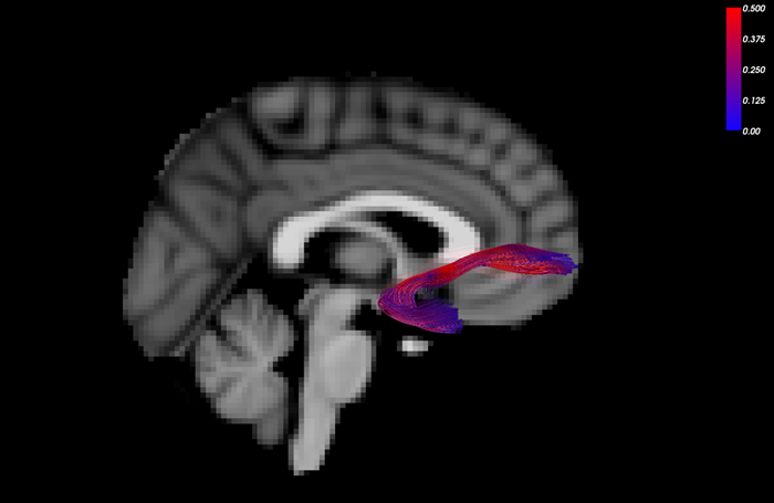 Tractography of Uncinate fasciculus in sagittal view, colored by fractional anisotropy (FA), blue denotes low FA, red high FA
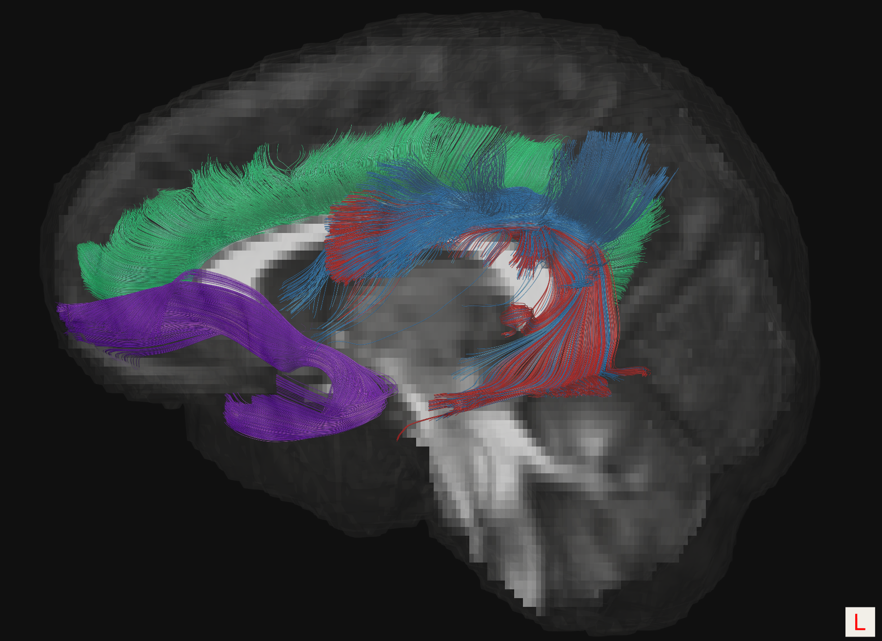 Tractography of a typically developing cingulum (green), superior longitudinal fasciculus (blue), arcuate fasciculus (red), and uncinate fasciculus (purple). Image from "Diffusion Tensor Imaging in Autism Spectrum Disorder: A Review" by Travers et al (2012).
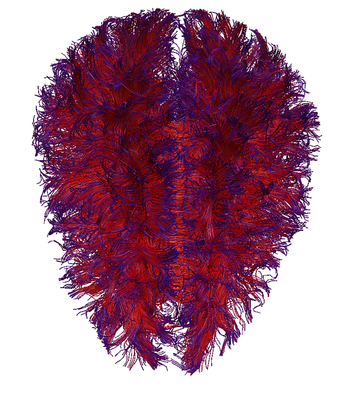 Whole brain tractography, colored by fractional anisotropy (FA), blue denotes low FA, red high FA
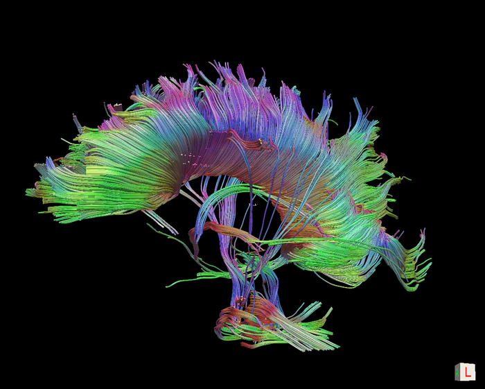 Colorful Corpus Callosum Tractography
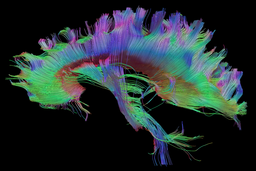 More colorful Corpus Callosum Tractography
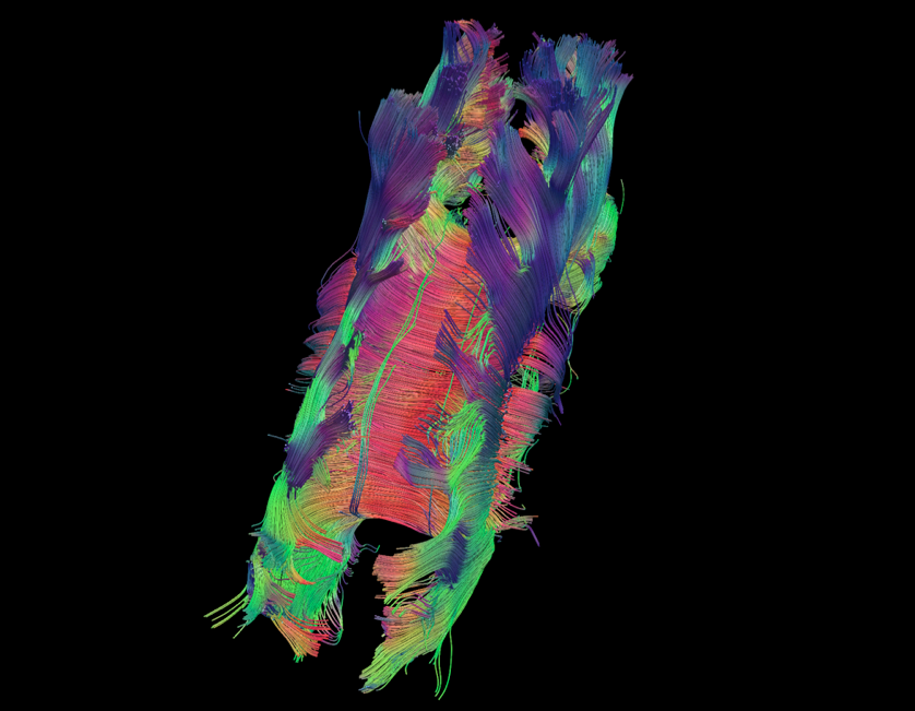 Colorful Corpus Callosum Tractography viewed from the top
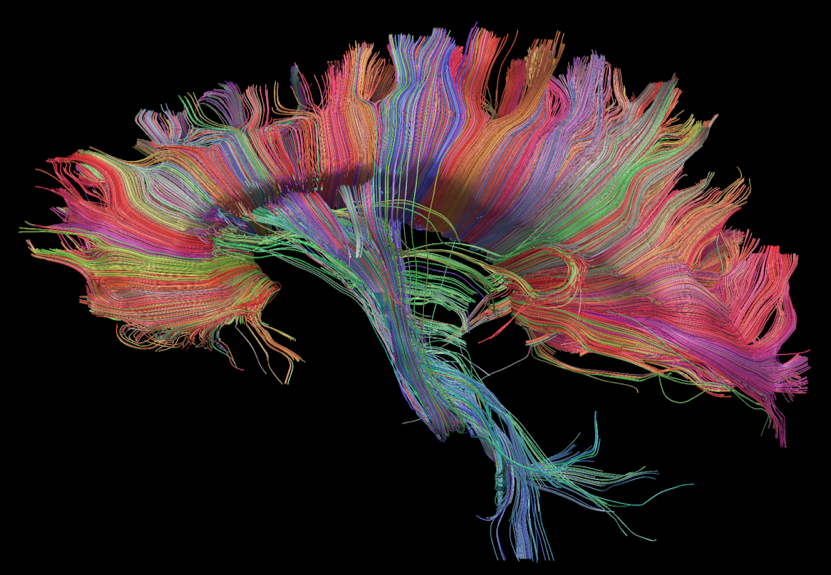 Colorful Corpus Callosum Tractography colored by end point location
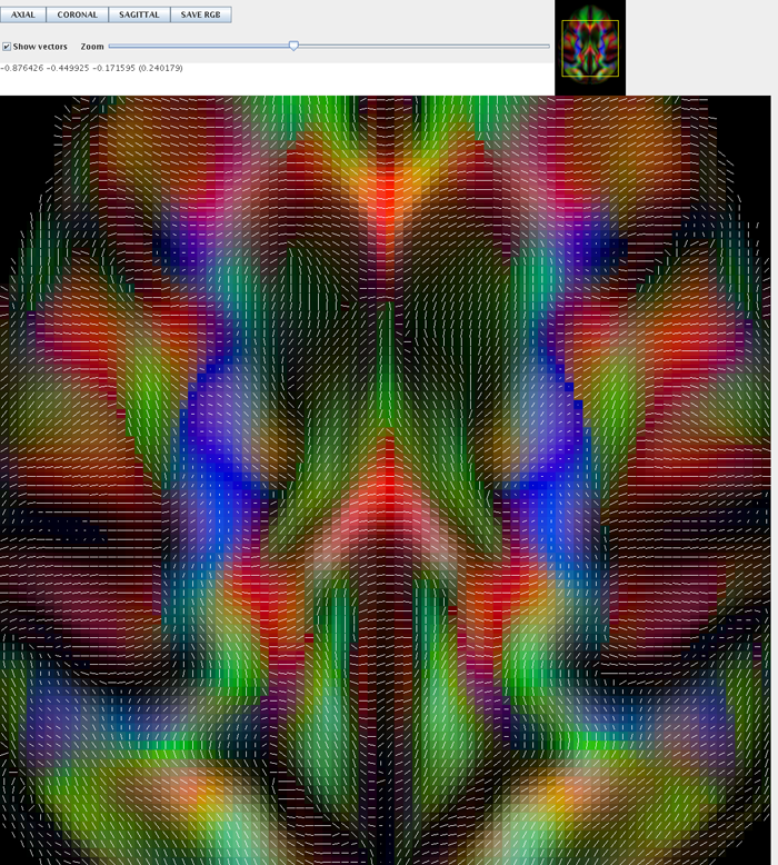 Axial view of tensor directions, with in the center in red the corpus callosum, in blue the superior corona radiata and in green the cingulum
 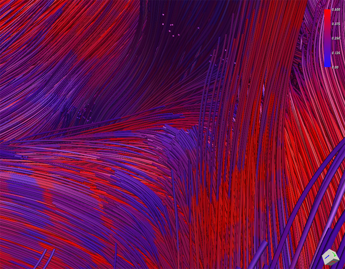 Inside the Corpus Callosum with Tractography
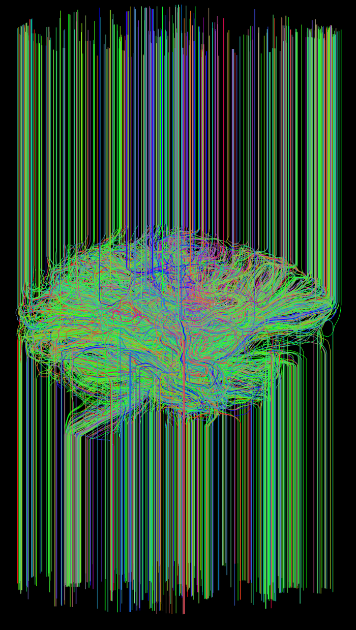 Oops, mistake Tractography