A Coregistration Challenge
This example shows how BrainSqueezer can coregister a particularly challenging case. A PET scan of a monkey must be matched to a T2-weighted MRI scan. The challenge is that, due to technical limitations, only the front half of the monkey's brain was scanned in the PET scanner. When first read into BrainSqueezer, the images look like this:
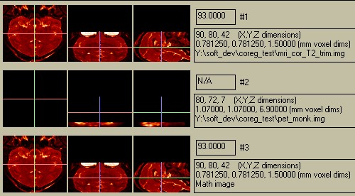
Figure 1. Original Display.
The PET image has only 7 slices in the Z-dimension (see file-size info to right of cenral row), so it appears much smaller than the MRI image. The PET value appears as "N/A" because the current location (crosshair) is not within the PET image. The anterior part of the brain is at the bottom of the PET image, and only half of the brain is shown. This would be a challenge for most automated coregistration packages! Let's start by getting the correct aspect ratio and pixel-to-pixel correspondence: click the "Coregister" button without changing any of the coregistration parameters. This reslices the Object (PET) volume into voxels having the same size as the Reference (MRI) image. We will also flip the PET image Anterior/Posterior by clicking on the middle "Up/Down" fllip button, to get the following display:
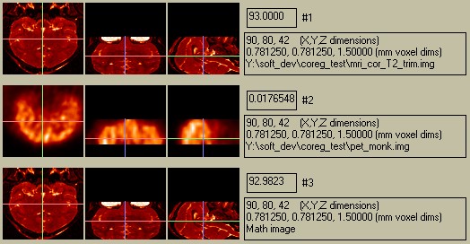
Figure 2. Resliced and Flipped Object image.
The PET data are displaced from the bottom. Thios will change the next time you click "Coregister"- don't let it alarm you! Notice that the voxel value now has a valid number for the PET image. However, the Math-Image is not very informative, since the difference in values is so great (~100 vs. ~0.5). We have three options for comparison:
We will go with option 3, and will no longer show the Math Image in this example. First we will set the contour value to show the outline of the head. Experience shows that setting the value on the histogram (go to the "Histogram" menu) as shown below is a good first guess:
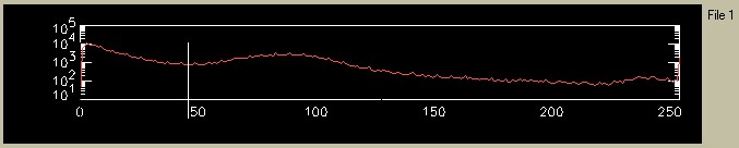
Figure 3. Setting the value for the MRI contour level.
After selecting "Contour: -> Ref Image" from the pulldown menu and increasing the zoom to 2, the images appear as follows:
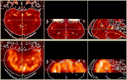
Figure 4. Contour based on the Refernce (MRI) image overlaid on itslef (top) and on the Object (PET) image at the bottom.
Already we are pretty close! Now we can tweak the coregistration parameters. If the pixel dimensions are accurate, we shouldn't have to change the zoom factors at all (since both images are from the same monkey). After a liesurely 60 seconds of changing the translation and rotation parameters, I arrived at the following alignment:
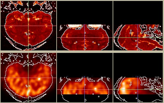
Figure 5. PET image (bottom) coregisterd to MRI image (top).
This looks pretty reasonable. If you disagree, you can change it yourself in a few seconds...To write the coregistered image to afile, click on "Write Object Image". A new image file will be created, named something like "pet_monk_coreg.img", and placed in the same directory as the original image (if you have write-permission). Note that the black region at the bottom of the central and right-hand PET images (the axial and sagittal slices) consists of 0's. These 0-valued voxels are included in the resliced Object volume and are also included in the written Object volume. This is usually the behavior you want e.g. to draw a ROI on the MRI image and overlay it onto the PET image.