Voxel-Based Morphometric
Approach to Modeling Stroke Lesions in Diffusion Weighted
Images
(c) 2013 Moo K. Chung, Dong-Eog Kim
mkchung@wisc.edu
Department of Biostatistics and Medical Informatics
Waisman Laboratory for Brain Imaging and Behavior
University of Wisconsin-Madison
Dongguk University Ilsan Hospital, Korea
Description
August 14, 2013
We
present a streamlined pipeline for quantifying a
collection of stroke lesion images in diffusion weighted images (DWI).
Although Bernoulli models are often used in modeling a
collection of binary images, the Bernoulli models
actually break down for testing statistical
significance of common overlaps. To remedy the
limitation of the Bernoulli models, we propose to
adapt the random field theory often used in
voxel-based morphometry(VBM) to stroke lesion images.
If you are using the Matlab codes/sample data below
for your publication, please reference [4] in the
Referenes. The codes have been tested under Matlab
R2009b 64bit version in Macbook OS X 10.6.8.
For bug reports, email
mkchung@wisc.edu
Stroke Lesions Binary Segmentaion Data
August 14, 2013
Stroke lesions are
semiautomatically segmented and saved as binary images
of size 370 by 301 in stroke.mat.
The first group (group1) consists of 58 subjects and the
second group (group2) consists of 23 subjects (Figure
1). For the detailed description of the
groups, see [4]. The Matlab script for the codes below
is given in stroke-v1.m
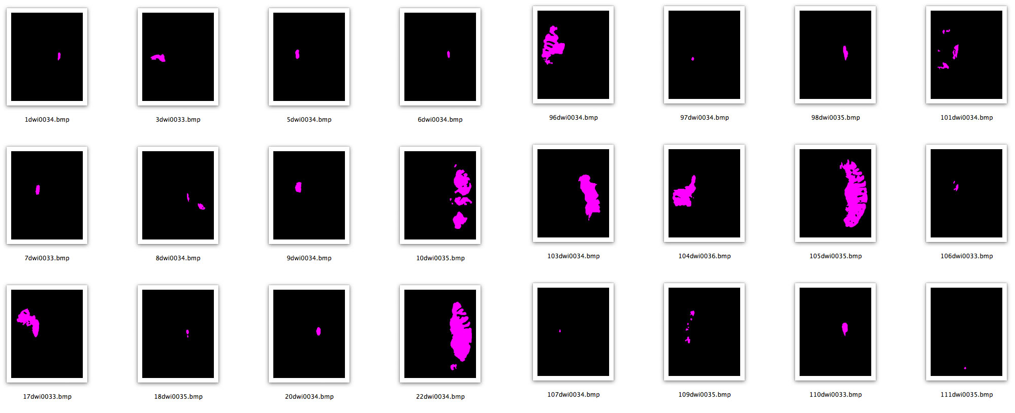
Figure 1. The
first four columns are images of
segmented infarct lesions in acute (less
than 7 days) stroke patients whose
dysphagia was improved after one month
(n = 58). The last
four columns are images of stroke
patients whose dysphagia was not
improved after one month (n = 23). We
are only showing a subset of the whole
data. We are interested in determining
if there is any common region where
stroke lesions consistently occur.
Voxel-Based Morphometry on Lesions
Segmentaion
August 14, 2013
We
perform the two sample test on the equality of the
proportions of stroke lesions (Figure 2):
[pvalue Zvalue] = stat_proportion(group1, group2);
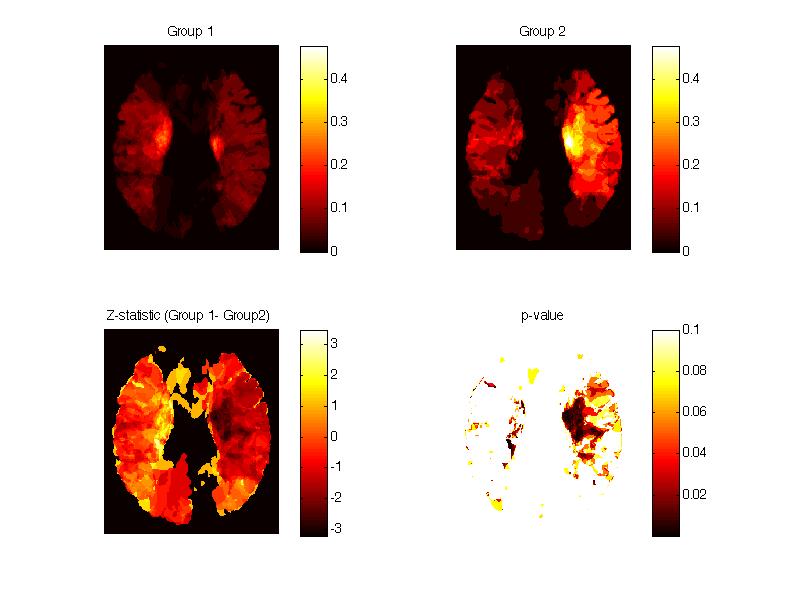
Figure 2. The
statistical significance of the equality
of the proportions of stroke lesions
between the two groups.
The problem with the above approach is the lack of
spatial smoothing that inflates a lot of small speckles
of false positives that are caused by discretization
errors. After smoothing with 10 pixelwide FWHM, we can
signifcantly redue such discretizatione artifacts (Figure
2). Smoothed version is done as follows.
FWHM=10;
group1smooth= stat_smooth(group1,
FWHM);
group2smooth=stat_smooth(group2, FWHM);
[pvalue Zvalue] = stat_proportion(group1smooth, group2smooth);
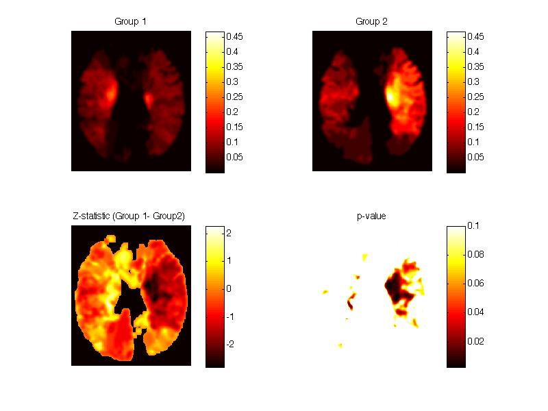
Figure 3. The
statistical significance of the equality
of the proportions of stroke lesions
between the two groups after Gaussian
kernel smoowhing with 10 pixelwide FWHM.
However, after
multiple comparsions correction based on the random
field theory, the signal in Figure 3 wil be
completely disappear (Figure 4). The
random field theory based multiple comparions will be
done using
pcorrected=stat_Zcorrected2D(Zvalue,
FWHM);
figure; imagesc(pcorrected); colormap('hot');
colorbar; caxis([0 0.1])
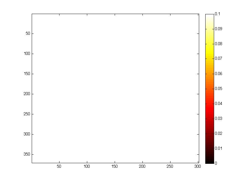
Figure
4. The
statistical significance
after the random field
theory based multiple
comparisons.
References on Voxel-Based Morphometry
August 14, 2013
- Chung, M.K.,
Dalton, K.M., Alexander, A.L., Davidson, R.J. 2004. Less
white
matter concentration in autism: 2D voxel-based morphometry.
NeuroImage
23:242-251.
- Chung, M.K.,
Shen, L., Dalton, K.M., Davidson, D.J. 2006. Multi-scale
voxel-based
morphometry via weighed spherical harmonic representation.
International Workshop on Medical Imaging and Augmented
Reality (MIAR).
Lecture Notes in Computer Science (LNCS). 4091:36-43.
- Oakes, T.R.,
Fox, A.S., Johnstone, T., Chung, M.K., Kalin, N., Davidson,
R.J. 2007. Integrating
VBM into the general linear model with voxelwise anatomical
covariates. NeuroImage
34:500-508.
- Chung, M.K.,
Woo, S.H., Lee, J. S., Kim, D.-E. 2013. Modeling
stroke lesions in diffusion weighted images using 2D random
field theory, SPIE Medical Imaging. submitted.